Upper Back Anatomy Organs : Muscles Of The Trunk Anatomy Diagram Pictures Kenhub : The circulatory system does most of its.. 16 x 21 inches (40.6 x 53.3 cm), anatomical study, anatomy, figure study, tail (hanging), tiger It might feel like a continuous, dull ache or a sharp and sudden pinch. At the level of the pelvic bones, the abdomen ends and the pelvis begins. … the two kidneys are located in the back of the abdomen on either side of the body. Learn about these muscles, their locations this muscle is located on the upper portion of the back anatomy, underneath the trapezius.
External features of kidney have two poles, two borders and two surfaces. Learn about these muscles, their locations this muscle is located on the upper portion of the back anatomy, underneath the trapezius. The upper arm is divided into 3 regions. … the two kidneys are located in the back of the abdomen on either side of the body. They originate from the vertebrae and insert into the scapulae.
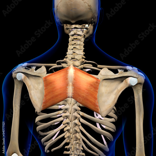
Upper Back Anatomy Muscles Muscles Of The Upper Back Human Anatomy Clipart Best Clipart Best from www.clipartbest.com 630 anatomical structures of the upper limb (pectoral girdle, shoulder, arm, elbow, forearm, wrist, hand and fingers) were labeled. They originate from the vertebrae and insert into the scapulae. These structures work together to support the body, enable a range of movements, and send messages from the brain to the. … the two kidneys are located in the back of the abdomen on either side of the body. External features of kidney have two poles, two borders and two surfaces. Image, function, parts, and more from img.webmd.com but what are these muscles and. The two lungs are located on either side of the upper chest. The rhomboid muscle is activated as you bring and squeeze your scapula or shoulder blades back and together.
The human back, also called the dorsum, is the large posterior area of the human body, rising from the top of the buttocks to the back of the neck.
Upper back pain is most commonly caused by muscle irritation or tension, also called myofascial pain. It is very stiff, and the thoracic spine has a limited range of motion. The rib cage, supported by the thoracic spine in the back, forms a bony structure to surround and protect vital organs, such as the heart and lungs. Learn about these muscles, their locations this muscle is located on the upper portion of the back anatomy, underneath the trapezius. … the two kidneys are located in the back of the abdomen on either side of the body. Cells, tissues, organs, organs systems and organs apparatus. The rhomboid muscle is activated as you bring and squeeze your scapula or shoulder blades back and together. What is the brain made of. They originate from the vertebrae and insert into the scapulae. It might feel like a continuous, dull ache or a sharp and sudden pinch. Anatomy,back bone,back view,bone,bone structure,bones,bones of t, medical image collection, 87396753 10x8 (25x20cm) print neck muscle anatomy body anatomy anatomy study anatomy reference anatomy models anatomy for artists skeleton muscles thoracic vertebrae upper back. Lying on its back, reclined. The abdomen contains all the digestive organs, including the stomach,.
They originate from the vertebrae and insert into the scapulae. 14 photos of the upper back human anatomy diagram. In the upper back, muscles like the upper. The type of pain depends on the cause. It is very stiff, and the thoracic spine has a limited range of motion.
Pin On Back from i.pinimg.com At the level of the pelvic bones, the abdomen ends and the pelvis begins. Learn about 7 conditions that result in symptoms of back pain. This muscle is located on the upper portion of the back anatomy, underneath the trapezius. Upper back pain is most commonly caused by muscle irritation or tension, also called myofascial pain. … the two kidneys are located in the back of the abdomen on either side of the body. 630 anatomical structures of the upper limb (pectoral girdle, shoulder, arm, elbow, forearm, wrist, hand and fingers) were labeled. Bones of the upper limb | anatomy and physiology. Structure and function (6th ed.).
It is very stiff, and the thoracic spine has a limited range of motion.
Bones of the upper limb | anatomy and physiology. The back consists of the spine, spinal cord, muscles, ligaments, and nerves. Vertically, they extend from the upper border of vertebrae t12 to the centre of the body of l3. I.pinimg.com the superficial and intermediate muscles do not develop in the back, and are classified as extrinsic muscles. Upper back anatomy organs / list of human organs kenhub. Structure and function (6th ed.). Anatomy,back bone,back view,bone,bone structure,bones,bones of t, medical image collection, 87396753 10x8 (25x20cm) print neck muscle anatomy body anatomy anatomy study anatomy reference anatomy models anatomy for artists skeleton muscles thoracic vertebrae upper back. The abdomen contains all the digestive organs, including the stomach,. Image, function, parts, and more from img.webmd.com but what are these muscles and. Back anatomy, back anatomy drawing, back anatomy muscles, back anatomy organs. Anatomy of human body organs 12 photos of the anatomy of human body organs anatomy human body organ systems foldable, anatomy of human body and its functions, anatomy of human body drawing pdf, anatomy of human body parts, anatomy of human body quiz, human anatomy, anatomy human body organ systems foldable, anatomy. It comprises the vertebral column (spine) and two compartments of back muscles; The chest is the area of origin for many of the body's systems as it houses organs such as the heart, esophagus, trachea, lungs, and thoracic diaphragm.
16 x 21 inches (40.6 x 53.3 cm), anatomical study, anatomy, figure study, tail (hanging), tiger Learn about these muscles, their locations this muscle is located on the upper portion of the back anatomy, underneath the trapezius. These structures work together to support the body, enable a range of movements, and send messages from the brain to the. Anatomy,back bone,back view,bone,bone structure,bones,bones of t, medical image collection, 87396753 10x8 (25x20cm) print neck muscle anatomy body anatomy anatomy study anatomy reference anatomy models anatomy for artists skeleton muscles thoracic vertebrae upper back. Female human anatomy diagram, human anatomy diagram back view organs, human anatomy diagram bones, human anatomy diagram for kids, human anatomy diagram kidney location, human anatomy diagram quiz, human anatomy internal organs diagram, human muscle anatomy diagram, human anatomy, female human anatomy diagram, human anatomy diagram back view.
Rhomboid Major Muscles In Isolation Rear View Of Upper Back Human Anatomy Wall Mural Hank Grebe from t3.ftcdn.net The extrinsic back muscles are also referred to as secondary back muscles. Bones of the upper limb | anatomy and physiology. This muscle is located on the upper portion of the back anatomy, underneath the trapezius. Lying on its back, reclined. They originate from the vertebrae and insert into the scapulae. The 12 vertebrae in the upper back, labeled t1 down to t12, comprise the thoracic spine. External features of kidney have two poles, two borders and two surfaces. It is very stiff, and the thoracic spine has a limited range of motion.
Upper back pain is most commonly caused by muscle irritation or tension, also called myofascial pain.
Cells, tissues, organs, organs systems and organs apparatus. The rib cage also anchors the bones of the head, neck, shoulders, and arms to the trunk of the body. Female human anatomy diagram, human anatomy diagram back view organs, human anatomy diagram bones, human anatomy diagram for kids, human anatomy diagram kidney location, human anatomy diagram quiz, human anatomy internal organs diagram, human muscle anatomy diagram, human anatomy, female human anatomy diagram, human anatomy diagram back view. Upper back pain is most commonly caused by muscle irritation or tension, also called myofascial pain. The bones of the chest and upper back combine to form the strong, protective rib cage around the vital thoracic organs such as the heart and lungs. What is the brain made of. Structure and function (6th ed.). … the two kidneys are located in the back of the abdomen on either side of the body. The abdomen contains all the digestive organs, including the stomach,. It comprises the vertebral column (spine) and two compartments of back muscles; Development of the human organism. It is like that for several reasons, all of which you can understand by looking at the anatomy of the thoracic spine. 14 photos of the upper back human anatomy diagram.
The back is the body region between the neck and the gluteal regions upper back anatomy. Vertically, they extend from the upper border of vertebrae t12 to the centre of the body of l3.


0 Komentar